Wednesday, November 13, 2013

National Diabetes Month: A Perfect Time to Schedule Your Annual Eye Exam

November is National Diabetes Month and a perfect reminder for individuals with diabetes to schedule their annual eye exam. It’s not just about controlling glucose and blood sugar levels - an annual screening for diabetic retinopathy is also incredibly important in keeping you healthy. 

Diabetes is the most common cause of blindness in adults under the age of 70, and diabetic retinopathy affects roughly 25% of diabetics. Diabetic retinopathy is caused by the deterioration of blood vessels that nourish the retina. When this happens, the vessels weaken and leak fluid that would have otherwise provided healthy nourishment for the retina. 

The early stages of the disease are quite subtle and most patients don’t notice a change in vision until a much later stage when they experience blurred vision or floaters in the eye. The longer the individual has had diabetes and the level of the individual’s diabetic control are both factors that can lead to a greater chance of developing diabetic retinopathy. 

If the disease is caught and monitored in an early stage, more of the sight-damaging effects can be slowed or stopped completely. 

Ophthalmologist Clayton Falknor, M.D. explained, “If a person with diabetes waits until they notice vision problems to have their eyes examined, they run a much greater chance of having more severe retinal disease which may require laser treatments, injectable medications, and possibly even surgery. Their primary care physician also needs to know the extent of retinopathy present to guide the aggressiveness of blood sugar management on a day-to-day basis.”

A comprehensive annual exam is one of the best ways to keep diabetic retinopathy in check. Make an appointment with one of our skilled ophthalmologists at the Eye Clinic of Austin to ensure early diagnosis and treatment.

Source: http://www.emaxhealth.com/1020/national-diabetes-month-good-time-focus-eye-health

Wednesday, November 6, 2013

Research Finds New Eye Drops Could Treat Macular Degeneration

New research published in the journal PLos One earlier this month has many sufferers of age-related macular degeneration (AMD) hopeful for the possibility of a new treatment. 

Age-related macular degeneration is an eye disease that gradually destroys the macula, the part of the eye that provides sharp, central vision needed for seeing objects clearly. According to the Centers for Disease Control and Prevention, almost 2 million Americans over the age of 40 have poor vision caused by AMD.

The new findings experimented with a chemical PPADS (short for pyridoxalphosphate-6-azophenyl-2’,4’-disulfonic acid) to repair AMD-related damage to the eye. Researchers at Tufts University in Massachusetts induced tissue damage and blood vessel growth characteristic of AMD in anesthetized mice and then applied PPADS daily, which resulted in the chemical healing the eye damage. 

The positive implication from this is the fact that a topical application of a drug - for example, in the form of an eye drop - could ultimately be used on humans to treat AMD. Previous research sought to show that certain dietary supplements, such as lutein, were effective in reducing the risk of progressing from dry macular degeneration to wet macular degeneration. This new study has many researchers excited for the possibilities of self-administered treatments. 

Ophthalmologist Thomas Henderson, M.D. explained, “If confirmed in humans, this chemical, given as an eye drop, could potentially reduce or eliminate the huge cost of and need for monthly injections into the eye to stabilize wet macular degeneration and preserve vision.”

Because this research is the first of its kind to demonstrate a topical application of a drug treating wet macular degeneration, much more research is now due in order to confirm this study’s findings.

Photo Credit: Ross Toro, myhealthnewsdaily.com 

Wednesday, October 30, 2013

Dangers of Colored Costume Contact Lenses

costume contact lenses
Those cat eye lenses really might make your costume stand out from the crowd, but don’t let decorative contact lenses play a trick on you this Halloween. Did you know that costume contact lenses require a prescription? Just like your regular contacts, costume lenses are considered to be medical devices and even with perfect vision, a prescription and eye exam is required.  Wearing lenses that aren’t properly fitted for you can scratch the eye and actually cause blood vessels to grow into the cornea. This could also lead to eye infection and potential permanent vision loss. A scary thought, indeed.

Selling non-prescription lenses, like costume lenses found online or in beauty shops, is illegal. Many consumers are unaware that these lenses are not FDA-approved and with advertising claims like “one size fits all” even the most savvy shopper might consider trying out a pair without realizing the importance of a proper prescription.

Ophthalmologist Clayton Falknor, M.D. offered, “If most people understood the potential for permanent vision loss by infectious keratitis that can occur with non-approved or ill-fitted contacts, very few would be willing to take the risk in using them.”

Whether colored contact lenses are a personal style choice or the finishing touch to the perfect Halloween costume, they should be fitted to your eye and purchased at a retailer that requires a prescription or from an eye care professional.


Wednesday, October 23, 2013

Wear Protective Eyewear During DIY Projects

October in Texas brings that cooler weather we all love and many begin work on DIY (do-it-yourself) home projects. People don’t realize how often injuries to the eye take place at home while not wearing protective eyewear. According to the American Academy of Ophthalmology, more than 2.5 million eye injuries occur in the US each year with 50,000 people permanently losing part or all of their vision.
Ophthalmologist Thomas Henderson, M.D. explained, “In my years taking emergency calls, I have seen many unfortunate and preventable injuries, some of which caused permanent, major loss of sight.  It always seems to be the ‘one time’ you did not wear the protective eye wear that causes these horrible things to happen.”
Many know eyewear is a must while mowing or trimming the lawn, but even the most mundane tasks require eye protection. Splashes from household cleaners, solvents or paints can also cause eye injury. Individuals who believe their own prescription glasses are enough protection are mistaken. In fact, wearing just your prescription glasses may cause further injury. Many safety goggles or glasses fit over regular glasses, and they absolutely should be worn.
Prevent Blindness America advises people wear protective eyewear approved by the American National Standards Institute (ANSI). This eyewear will have a “Z-87” logo on the frames. For additional information about eye safety at home, visit preventblindness.org/eye-safety-home.

Wednesday, October 16, 2013

Blood Test Hopes to Improve Treatment for Children with Cataracts

blood test for children with cataracts
Researchers in the UK have developed a new blood test, which hopes to improve diagnosis and treatment for children born with congenital cataracts.

Diagnosing the presence of a cataract is fairly easy, the hard part is determining the cause of the child’s cataract. There are more than 100 different mutations linked to congenital cataracts and with no family history of cataracts, diagnosis of cause can be very difficult. Determining this cause is important because the child’s cataract may have formed due to another congenital condition such as a metabolic disorder or Down syndrome. In approximately 25 percent of children with cataracts at birth, the condition is due to another genetic cause.

The blood test developed analyzes every known DNA mutation that causes cataracts to help speed up diagnosis of the cause. This leads to better treatment and the ability to inform families of genetic risks for the future.

Ophthalmologist Clayton Falknor, M.D. offered, “Treatment for children with cataracts is tailored to the child. In mild cases, no removal is necessary, but in many cases, the cataract must be removed in order for the child to develop his or her best possible eyesight. Depending the child’s age and other factors, a lens implant may be placed at the time of surgery to replace the clouded lens.”

A cataract is a clouding of the lens in the eye that affects vision. In the US, only 0.4 percent of children are born with cataracts.

Sources: 

http://www.bbc.co.uk/news/health-24084614
http://www.childrensnational.org/DepartmentsandPrograms/default.aspx?Type=Dept&Id=345&Name=Ophthalmology&SubType=ConditionOrTreatment&SubId=485&subname=Cataract%20and%20other%20lens%20abnormalities

Wednesday, October 9, 2013

Smartphone Advances Provide Mobile Eye Exams in Third World Countries

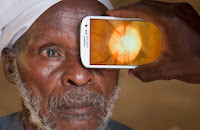
Peek iphone app
With everyone in the US abuzz about the new iPhone and Apple operating systems, other smartphone advancements are making an even bigger impact worldwide. One of the biggest causes of blindness around the world is due to untreated cataracts and uncorrected refractive errors. Because traditional exam equipment is immobile and very expensive, people in remote rural areas of third world countries rarely receive eye exams or eye care. This is now beginning to change, however, due to advancements with the smartphone and smartphone applications (apps).

A new smartphone app called Peek is revolutionizing the way eye exams are provided in these countries. Using a smartphone and an external clip-on device, Peek can check for cataracts, perform simple vision tests and scan the retina for disease, allowing conditions such as glaucoma, macular degeneration, and diabetic retinopathy to be diagnosed.

Minimal training is required to operate Peek as the information is gathered and then sent to experts around the world for diagnosis. In addition, the GPS data the smartphone and app gather is also very helpful because this information allows for follow-ups and helps other health organizations better target mass treatment campaigns.

Ophthalmologist Thomas Henderson, M.D. offered, “This iPhone application represents a new pathway to bring improved eye care to many people throughout the world. In the United States, I foresee its potential use in emergency room consultations with a physician who is not on site.”

Peek is currently being tested in Kenya, and their team will publish results at the end of their trials in early 2014.

Source: http://singularityhub.com/2013/09/06/will-peeks-mobile-eye-exam-system-take-a-bite-out-of-developing-world-blindness/

Wednesday, September 25, 2013

Artificial Corneas Prove Promising for Transplants

A recent Swedish study proves artificial corneas may be a promising alternative to donor corneas. The study involved 10 patients with corneal damage and significant vision loss between the ages of 18 and 75. These individuals were the first in the world to be fitted with biosynthetic tissue as opposed to human donor corneas. Two years following the surgery, the patients had not experienced any serious reactions or complications.

Corneal transplants can be necessary in conditions such as kerotoconus (corneal damage where there is gradual thinning and bulging of the cornea). Disease of the cornea is one of the major causes of blindness worldwide and is often due to trauma or infection. In many parts of the world, there is a shortage of corneal donors causing waiting lists for the surgery.

According to Ophthalmologist Clayton Falknor, M.D., “A shortage of corneal donors is not typically an issue in the United States due to the excellent eye bank system. We are normally able to secure a donor cornea as necessary. However, globally this is a serious issue and research into synthetic alternatives is very important.”

Initial results from the Swedish study show that biosynthetic corneal implants could offer a solution to the current donor shortage worldwide. However, as the first study of its kind, far more research and much larger numbers of patients is needed to confirm the safe use of biosynthetic corneas.


SOURCE:
http://www.shorehamherald.co.uk/news/top-stories/health/partial-success-of-artificial-corneas-1-984091

Wednesday, September 18, 2013

Safety: LASIK VS Contact Lenses

lasik safety vs contact lenses
When considering LASIK over contact lenses, many patients’ biggest concern is the safety of LASIK. What many do not realize is that patients have a higher risk of losing correctible vision with contact lenses than by undergoing LASIK eye surgery, a one-time surgical procedure. In fact, that risk is perhaps up to 10 times higher.

Many contact lens wearers do not properly take care of their lenses and this is what can cause infection. For example, wearing contacts during sleep, swimming in them, not cleaning them properly, and not replacing them regularly increases the risk of infection and could damage eyesight permanently. However with LASIK, the laser technology being used today is the safest it has ever been.
LASIK provides phenomenal precision and very predictable results. A person with a typical refractive error has a 93% chance of having their vision fully corrected after the LASIK procedure and without requiring additional treatment, contacts, or glasses. For those who still have some refractive error after surgery, the majority can be re-treated to achieve optimal results. In terms of the risk of losing correctible vision as a result of the surgery, the risk is extremely small – only 1 in 1,000 experience significant complications. 

Ophthalmologist Clayton Falknor, M.D. said, “Of all the things that I do surgically, LASIK is the most accurate and the most consistent with the best results. After healing, your eyesight is normally stable for 10-20 years, depending on your age at the time of surgery.”

If you are interested in LASIK, but concerned about the safety, speak with your ophthalmologist about the procedure. At Eye Clinic of Austin, we offer free LASIK consultations to determine candidacy for the procedure. This appointment also provides patients an opportunity to speak with the eye doctor, ask questions, and discuss any concerns.

Wednesday, September 11, 2013

Study Ties Cataract Surgery to Longer Life

An Australian study published this month in Ophthalmology, the journal of the American Academy of Ophthalmology, shows that people with vision loss attributed to cataracts who have had cataract surgery live longer than those with cataract-related vision loss who forgo the procedure. Those who had cataract surgery were found to have a 40 percent lower long-term mortality risk.

From 1992 to 2007, the study followed 354 individuals aged 49 years and older who had been diagnosed with vision impairment due to cataracts. Adjustments were made for a number of mortality risk factors including age, gender, diabetes, smoking, body mass index, hypertension, measures of frailty, and comorbid disease.

Corroborating previous research indicating that individuals with visual impairment were likely to have a greater mortality risk than their peers with normal vision, this study continues to suggest that healthy vision results in general good health.

The correlation between visual impairment correction and reduced mortality is not completely clear, but possible factors include improved physical and emotional well-being, greater optimism and confidence, and ability to comply with prescription medications.

Ophthalmologist Thomas Henderson, M.D., explained, “My experience matches the results of this study. Even with Alzheimer’s, the patients who get their cataracts removed do better because of increased contact with the world. Thirty years ago, I had a patient who was bilaterally blind from cataract. She was on psychiatric medication because she thought people were stealing from her and others thought she was just paranoid. I did cataract surgery giving her normal vision in both eyes. She stopped her medication because she was better oriented to the world, and the stealing stopped because she could see.”

A cataract is a clouding of the lens in the eye that affects vision. Most cataracts are related to aging and by age 80, more than half of all Americans either have a cataract or have had cataract surgery. To determine if you have cataracts, contact your ophthalmologist and schedule a comprehensive dilated eye exam.

Wednesday, September 4, 2013

Allergies VS Dry Eye Syndrome

If eyes are uncomfortable, scratchy or
gritty, these are signs that dry eye
may be the cause
Two of the most common forms of eye irritation are eye allergies and dry eye syndrome (frequently called dry eye). Both can cause itching, and many people mistake dry eye symptoms for allergies. If eyes are uncomfortable, scratchy or gritty, these are signs that dry eye may be the cause.

Dry eye occurs when eyes do not produce the right quantity or quality of tears. It is among the least understood eye conditions and affects the largest number of people, up to 20% of the population. If left untreated, the risk of permanent damage and scarring to the front of the eye increases.

Dry eye is most common in women older than 50 due to hormonal changes, and only an eye doctor can diagnose the issue. Typically, the doctor will perform one or more tests to assess the condition. Ophthalmologist Clayton Falknor, M.D. explained, “At Eye Clinic of Austin, we use the TearLab® Osmolarity Test. This gives us information about the concentration of salt in a patient’s tears, and helps us diagnose the severity of the issue.”

Initial treatment of mild dry eye consists of lubricating the eye with topical artificial tears or possibly a prescription eye drop called Restasis.

“If Restasis is used properly twice a day for six months, the result is typically dramatic improvement for 90% of patients. This prescription medication can significantly improve quality of life,” said Ophthalmologist Thomas Henderson, M.D.

To determine if you have dry eye syndrome, contact your eye doctor and schedule a comprehensive eye exam.

Thursday, August 29, 2013

New Study Ties Early Menopause to Glaucoma

Women who experience early meno-
pause may be at higher risk for dev-
eloping glaucoma later in life.
A new study from the Netherlands Ophthalmic Research Institute in Amsterdam shows women who experience early menopause may be at a higher risk of developing glaucoma later in life. The study tracked 3,000 women, and those who went through natural menopause before the age of 45 were more than twice as likely to be diagnosed with open-angle glaucoma than women who went through menopause at age 50 or older.

Glaucoma is caused by fluid accumulating in the eye, which then puts pressure on the eye’s optic nerve. It is a leading cause of blindness in the US, and has no symptoms or pain when it first develops. Medicine, laser treatment, and conventional surgery are used to treat the disease. Ophthalmologist Clayton Falknor, M.D. explained, “While these treatments can help prevent future loss of vision, they do not improve sight already lost from the disease which is why it’s so important to schedule eye exams as you age.”

This study’s result signals that female hormones may protect against the disease. Hormone replacement therapy is thought to reduce fluid pressure in the eye, and researchers of the study also note that hormone levels rise during pregnancy causing fluid pressure in the eye to decrease.

“This study shows the potential value of analysis of large amounts of data. Although I have seen patients for over 30 years, this is not a connection that I have considered. At this time, I am not prepared to prescribe hormone therapy for menopausal women with glaucoma,” said Ophthalmologist Thomas Henderson M.D.

As glaucoma research continues, it’s still very important to schedule comprehensive eye exams as you age to detect any issues and begin treatment, if needed.

Sources:
http://www.tele-management.ca/2013/08/study-links-early-menopause-to-glaucoma-risk/
http://www.nei.nih.gov/health/glaucoma/glaucoma_facts.asp#a

Wednesday, August 21, 2013

What to Do at What Age for Your Eyes

national eye exam month
August is National Eye Exam month and although each individual’s eyes are different, there are recommended times in your life in which to receive an eye exam. Here’s a summary of what to do at what age for your eye care:

INFANTS: All babies should receive an eye exam soon after birth. It’s also recommended to receive another exam between ages 6 to 12 months.

PRESCHOOL: At age 3, it’s important for children to receive a visual acuity test. The test results can show lazy eye or refractive errors, like nearsightedness, farsightedness or astigmatism.

SCHOOL AGE: Upon entering the 1st grade, another eye exam should be performed. As your child gets older, if any suspected vision issues or problems come up, then another exam should be performed. In this age group, nearsightedness is most common which can be corrected with eyeglasses or contact lenses, depending on the child’s age.

TEENAGERS & EARLY 20s: Teenagers with corrective lenses should receive an annual eye exam in order to determine changes in prescription and to detect any other issues. By the time people reach their early 20’s, vision development will typically remain steady. For this age group it’s also important to remember to protect eyes during activities and playing sports.

ADULTS 25 to 40: Vision changes little at this age, so this is the recommended time for LASIK eye surgery. For those without prescription lenses or a family history of eye disease, it’s best to receive at least one complete eye exam in your 20s and two complete exams in your 30s. During pregnancy, women may have vision fluctuations.

ADULTS 40 to 65: This is a time when many eye diseases may begin to emerge. At age 40, a comprehensive eye exam should be scheduled to check for any signs of age-related macular degeneration, cataracts, glaucoma and other problems. You should continue to schedule exams every two years or more often based on feedback from your ophthalmologist about any problems your eyes have.

65 AND UP: By age 65, 1 in 3 Americans will have a vision-impairing eye disease such as glaucoma and cataracts. Preventive eye exams should be an important annual appointment to help find any issues or continue treatment of any known problems as your ophthalmologist recommends. Vision may be saved or prolonged if eye disease is detected early on.

Source: American Academy of Ophthalmology

Wednesday, August 14, 2013

Stem Cell Therapy for Macular Degeneration to Be Tested

macular degenerationResearchers in Japan began recruiting patients on August 1 for a pilot study to determine the possibility of using induced pluripotent stem cell (iPSC) therapy to treat wet-type age-related macular degeneration.

Age-related macular degeneration is a common eye condition for individuals age 50 and older. There are two forms of the disease, dry and wet. The wet form is more severe and is defined by damage to the retinal pigment epithelium (RPE), cells located in the back of the eye.

“Macular degeneration is the leading cause of vision loss in older adults. Currently, there is no way to cure the disease, only slow its progression,” explained ophthalmologist Clayton Falknor, M.D.

In this study, the damaged RPE is removed and researchers then transplant the iSPC-derived RPE cell sheets into the affected area of the eye. Replacing the RPE cells may repair past damage to the patient’s eye, but the issue is finding and using RPE cells that the patient’s immune system does not reject.

Ophthalmologist Thomas Henderson, M.D. said, “If this works as hoped without significant negative side effects, it will revolutionize the therapy of wet macular degeneration.”

If your family has a history of macular degeneration or you believe may have macular degeneration, contact your eye doctor and schedule a comprehensive eye exam. Although research continues in macular degeneration, the current treatment is still slowing the disease and the sooner it can be detected, the bigger impact treatment can have.


SOURCES:
http://www.asianscientist.com/health-medicine/stem-cell-therapy-eye-disease-riken-japan-2013/
http://www.nei.nih.gov/health/maculardegen/armd_facts.asp#1

Wednesday, August 7, 2013

Laser Cataract Surgery Serves Baby Boomers Well

As the baby boomer population begins to age, so too do their eyes. Now in their late 50’s and into their 60’s, many are developing cataracts, a common occurrence which affects 50% of the U.S. population by age 80.

“What we are finding is baby boomers with cataracts are typically still employed, and they want to return to work as quickly as possible,” explained ophthalmologist Thomas Henderson, M.D. “Laser cataract surgery provides not only greater precision, but also quick recovery for patients. Many patients comment on how soon they were able to return to life as normal.”

Cataracts are the leading cause of blindness worldwide. A cataract is a clouding of the lens in the eye that affects vision. Symptoms of cataracts may include cloudy or blurry vision, colors seem faded, glare, a halo may appear around lights, poor night vision, double vision, and frequent prescription changes in your eyeglasses or contact lenses.


Normal Vision
Vision with Cataract


Ophthalmologist Clayton Falknor, M.D. offered, “Baby boomers are more interested than prior generations in achieving partial or complete glasses independence after cataract surgery. With the benefits of laser cataract surgery and advances in lens implant technologies, such as toric lenses for astigmatism management and multifocal lenses for reduced need for reading glasses, this outcome is increasingly encountered.”

To determine if you have cataracts, you should contact your eye doctor and schedule a comprehensive dilated eye exam. In addition to cataracts, your eye doctor can check for other age-related issues such as macular degeneration and glaucoma. Early detection and treatment for eye diseases may save your sight.

SOURCE: http://www.nei.nih.gov/health/cataract/cataract_facts.asp 

Monday, August 5, 2013

Treat Inflamed Eyelids Quickly to Avoid Future Issues

A red, inflamed eyelid could be caused by an infection at the root of the eyelashes – that is a stye. More commonly though, a red, swollen eyelid is caused by an infection of the oil gland in the cartilage plate that reinforces the back of the eyelid. This is called a chalazion. Chalazions are much more common than styes – perhaps 10 times more common.

The initial treatment for a stye or a chalazion is heat for 10 minutes, four times a day, for two days. If the problem is not resolved in two days and especially if it is a chalazion, it needs intense antibiotic treatment to kill the infection quickly.

A stye will disappear on its own if antibiotics are started by day 3, but chalazions will form scar tissue in the cartilage plate within the first five days. Then there will be a lump present on the lid for many months, perhaps even a year or more. Once the scar tissue forms, the only treatment is to cut out the scar tissue. It is far better to treat early if you do not want an ugly lump and a minor office surgery.

If you have a growth or swelling of any kind on your eyelids, make an appointment with your ophthalmologist.

Monday, July 29, 2013

Sun Exposure to Eyes Can Cause Potential Damage

Some research indicates sun exposure has the potential for damage to the lens and retina of the eye.

Aside from that, the white of the eye can be affected as well. The most common issues are pinguecula, pterygium and, more rarely, cancer.

Pinguecula may appear as a raised yellowish or grayish bump on the inner or outer side of the white of the eye. It may occasionally be irritated or dry out and make contact lens wear less comfortable. In and of themselves, they are not dangerous and we can treat the inflammation and drying effectively with topical eye drops. The only way to remove them is surgically, which is rarely necessary unless they become a concern cosmetically.

Pterygium is another benign growth on the inner and outer whites of the eye. They are vascularized growths that creep onto the colored portion of the eye. Pterygium may affect vision by inducing astigmatism or, if quite large, may even block vision directly. They also can be easily irritated and surgery is necessary to remove them.

Pinguecula and Pterygium are most common in people who spend a lot of time outdoors. Wearing good sunglasses with UVA, B and C protection is important to prevent them. If you develop a growth on your eye, make an appointment with your eye doctor.

Monday, July 22, 2013

Pupil Dilation Provides a More Accurate Prescription

If you have ever noticed that you can see more looking through an open doorway than you can see looking through the door’s peephole, you will understand why we dilate pupils.

When the pupil is small, we cannot see the details of the lens and the view of the retina is compromised. When we cannot see, we cannot adequately follow the progress of conditions such as diabetes, cataracts, macular degeneration, and the risk of retinal tears and detachments in the peripheral retina and we may miss important details that could lead to loss of vision.

We also dilate to understand a potential source of error in eyeglass prescriptions. Individuals with astigmatism or who are far-sighted tend to overfocus. When we dilate the pupil, we can relax that tendency and get a more accurate and comfortable prescription for eyeglasses.

We do not have anything to replace pupil dilation completely, but we have a partial substitute with the OPTOMAP — photography that gives a 210-degree view of the inside of the eye or 80% of the retina. This allows us to see somethings in better detail than a dilated exam, but there is still 20% we cannot see. At least every other year, we still need to dilate to look at the details of the lens that the OPTOMAP cannot see.

It should be noted that OPTOMAP, when used to avoid dilation, is not covered by insurance and you may pay out of pocket for the convenience of no dilation. In summary, though it may be inconvenient and a bit uncomfortable, dilation allows us to take better care of your eyes.

Wednesday, July 10, 2013

The Right Time to Remove Cataracts

Historically, a cataract was ready to remove when it was “ripe.” This antiquated term was used to delay surgery until the cataract was advanced. Thankfully, that era has passed. Today, wonderful improvements in both cataract surgery and lens implants, including laser cataract surgery, routinely provide excellent vision, (often without glasses, depending upon the implant).

Medicare pays for cataract surgery when vision is 20/40 or worse. If cataracts interfere with something important, such as driving or reading, Medicare may pay for surgery earlier. The key is that the symptoms must be caused by the cataract, interfere with important life activities and must not be correctable by simpler means such as new glasses.

If your vision is worse than 20/40 but meets your needs, a delay in cataract surgery usually will not harm your eye or make the surgery more difficult. Medicare has recently found that cataract surgery by itself reduces the risk of falling and breaking a hip by 20%. Do not wait so long that your cataract is visible or “ripe” in the old sense and risk breaking a hip. Instead, see your eye doctor regularly to monitor your cataract, and when the time is right, enjoy your best possible vision for the rest of your life.

Wednesday, June 26, 2013

Cataract Facts & Factors

The lens inside your eye is normally clear at birth and leaves the pupil dark. The lens gradually loses its transparency as we age. The increasing whiteness of the lens, visible in the pupil in advanced cases, is a cataract.

There is a genetic component to cataract formation, and there clearly is an aging component. Extensive exposure to sunlight, smoking, poor diets, and disease, such as diabetes, play a role in the growth of cataracts.

However, there are factors that might help prevent a cataract or at least slow its growth.

It is not practical to try to choose your parents or not get older. But, it would be good to wear a hat and/or sunglasses outdoors, eat an ideal diet, and consider vitamin and antioxidant supplementation. Also, do not smoke or drink to excess and do control any other chronic diseases well, especially diabetes.

Most people as they age have some degree of cataract and many of these limit vision and lifestyle by creating significant glare when driving or loss of fine detail at distance or near. Fortunately, cataract surgery can restore excellent visual function and, depending upon the options chosen, can reduce the need to wear glasses at distance or near.

If you are concerned that you may have cataract, seek an evaluation by an ophthalmologist who offers the latest options. At the Eye Clinic of Austin, we seek to offer you the options that we believe will be best you and for your vision—the same that we would choose for our family or ourselves in similar circumstances.

Monday, June 24, 2013

Are Generic Eye Drops the Same as Branded Eye Drops?

The short answer is: They have the same main ingredient in the same amount and concentration. The long answer adds: Nothing else in the bottle needs to be the same. So they might be equally effective as the branded, nearly as effective, or less effective than the branded.

It's much like comparing two different brands of vanilla ice cream. To be vanilla ice cream, it must contain vanilla. But every brand has a different recipe leading to subtle changes in texture and taste. Those subtle differences might affect the most important measurement: how much you like that ice cream. Likely, you have a real favorite, maybe Blue Bell, not just any generic vanilla ice cream.

Branded eye drops must go through a very expensive FDA approval process to get on the market. The reason the generics are less expensive is that they are required only to have the same concentration of the active ingredient. They do not have to prove they are as effective as the brand name. This makes for a difference in price and probable differences in effectiveness. Less effect is important in many patients and critical in some. Because neither you nor I have control over which generic is dispensed to you, control of your problem may vary over time.

Sometimes any vanilla ice cream is OK, but usually your favorite is best. Cost is important; so is proven effectiveness. Choose wisely. If you're experiencing chronic dry eye though, then make an appointment with your ophthalmologist to determine if you have dry eye disease.

Monday, June 17, 2013

What is Laser Cataract Surgery?

For more than 15 years, a few doctors have told their patients they were having laser cataract surgery. This was a simplification to avoid explaining the complex nature of the ultrasonic vacuum device used to remove the cataract. It was not a laser.

The Eye Clinic of Austin uses the LenSx femtosecond laser in cataract surgery. This allows us to make precise and reproducible incisions in the cornea, both to enter the eye and to reduce astigmatism; as well as inside the eye to open the lens capsule and break the lens into pieces for removal.

Surgeons have good hands, but no matter how good those hands are, the laser is more precise. This degree of precision leads to more predictable results from cataract surgery because it reduces some of the variables. The laser lens fragmentation reduces the amount of ultrasonic energy required to remove the cataract with phacoemulsification, which reduces the general irritation to the eye. Both of these improvements lead to better results and an increased chance that you will get the results you and your surgeon want.

If you need cataract surgery, contact the Eye Clinic Austin for your laser cataract surgeon.

Monday, June 10, 2013

What's Causing the Flashes of Light in Eyes?

The three most common explanations for seeing flashes of light include:

1. An ocular migraine.
2. A problem with the retina such as a tear or detachment.
3. A separation of the jelly-like fluid away from the retina called a Vitreous Detachment.

Most ocular migraines involve seeing a shimmering pattern from both eyes. This will last from 10 minutes to 30 minutes and then resolve.

With a retinal tear, sparks of light will be seen and will persist for a long time along with the sudden appearance of “floaters” or little black specks in the vision. Patients sometimes describe this as seeing a gnat or a spider web. It can be a line or a dot. Retinal tears are serious. They should be diagnosed and treated immediately to prevent a retinal detachment.

In the majority of cases, however, a person experiencing flashes of light has a condition that is not dangerous called Posterior Vitreous Detachment. In this, the jelly-like fluid in the back of the eye separates from the retina. This leads to a retinal tear in less than 2 percent of cases. Most flashing will stop, and floaters will diminish slowly.

The only way to know which condition applies to you is to have an exam with an eye specialist immediately.

Monday, June 3, 2013

Why You Should Consider LASIK

Austin is home to active residents who enjoy the outdoors: Biking, boating, camping, tennis, golf and horseback riding are all popular ways to enjoy all Central Texas has to offer. If you find contact lenses and glasses interfering with your outdoors fun, you might want to consider LASIK.

Of all the things that I do surgically, LASIK is the most accurate and the most consistent with the best results. Though some people are concerned about the cost of LASIK, it is less than the lifetime cost of glasses or contact lenses, and financing is available. After healing, your eyesight is normally stable for 10-20 years, depending on your age at the time of surgery.

We are still not able to make the cornea optically perfect, but we can consistently make your vision better than with glasses or contact lens correction. After surgery, more than 90 percent of patients will see 20 /20. The remaining 10 percent will see 20 /24 to 20 /40 and may opt for a touch-up procedure at no additional cost to get to 20/ 20.

LASIK addresses near-sightedness, astigmatism and far-sightedness. So if you are looking forward to a summer of outdoor activities on the lake, the tennis court or the links and find your contacts or glasses get in the way, consider LASIK surgery soon.

Tuesday, May 28, 2013

The Duties of a Doctor

When I attended my 40th reunion of my medical school class two weeks ago, I was reminded again in a most powerful and succinct way what it means to be a doctor.

Dr. Dan Foster, a world-renowned endocrinologist and teacher of medicine, gave a brief lecture about the ethics of medicine. In it he said that medical doctors have four duties. First, be competent. Second, cure disease and prevent it whenever possible. Third, treat symptoms when there is no cure. Fourth, comfort always.

We are committed to continually seeking new and better ways to cure and treat disease. The technological revolution in health care has brought the ability to see structures of the eye so clearly that we can detect disease at earlier stages than ever before. High-tech equipment like Ocular Coherence Tomography (OCT) wasn’t available 20 years ago and now I cannot imagine practicing without it. The duty of comfort was paraphrased to me once as: “Doc, they don’t care how much you know until they know how much you care.” And it remains true today.

It was wonderful to be brought to ponder the privileged and special relationship the physician has with his patients. I am so grateful to have the lessons learned as a medical student brought so clearly and forcefully forward into my life again.

Monday, May 20, 2013

When Red & Itchy Eyes May Be a Sign of Conjunctivitis

Red and itchy eyes may or may not be a sign of conjunctivitis – sometimes called “pink eye.” Conjunctivitis is an inflammation or infection of the thin tissue that covers the white portion of the eye (the “conjunctiva”) and lining of the eyelids. There are three types of conjunctivitis: viral, bacterial, and allergic.

Viral conjunctivitis is extremely itchy. It is not treated with medication, because it will run a definite and limited course without drugs. Bacterial conjunctivitis, on the other hand, improves with medication and will respond to antibiotics after three to four days. A warm, wet cloth held up to the affected eye for a few minutes may ease discomfort.

Both of these types of conjunctivitis ordinarily cause discharge and both are contagious.

Allergic conjunctivitis is an immune response to a foreign substance in the eye, such as pollen, mold, or dust. Another possible trigger may be contact lens wear, which causes “giant papillary conjunctivitis.” We treat most of these cases by suppressing the immune response with antihistamines or, in more severe cases, topical steroids. We ease discomfort with the use of artificial tears (which lubricate the eye) or the application of a cool, wet cloth. Allergic conjunctivitis is not contagious.

If your eyes are red and itchy, first see your eye doctor to determine whether or not you are contagious. Your doctor will tell you if you need a prescription and when you may return to work or school. Your doctor also will check to see whether your red eyes are a symptom of a more serious eye condition.

Monday, May 13, 2013

Certain Lens Technology Allows Advantages with Outdoor Activities

This beautiful spring weather has provided many of our patients with extra opportunities to enjoy (and sometimes become frustrated) by their favorite outdoor activities. Current lens technology allows multiple solutions for these challenges. 

We find that golf enthusiasts, especially those with decreasing midrange and near vision, need special progressive lenses. Those lenses maximize their peripheral and distance vision while providing clear vision at the tee. 

There are multiple spectacle and contact lens designs available that meet these requirements. Some golfers prefer polarized sun lenses, while others find these cause distortion in their peripheral field and prefer traditional tinted lenses in shades of amber and brown. Amber and brown lenses highlight subtle changes in topography on the green, helping the golfer to anticipate the path the ball will follow to the tee.
 
Monovision, a method of correcting one eye for distance and the other for near with contact lenses, may work well for a concert violinist, but not at all for a tennis or golf player who needs excellent depth perception. Creative flexible solutions are sometimes necessary. Great tools for maximum flexibility are press-on adds or clip-on sunglasses in multiple color tints. Gray for running in bright sunlight, polarized for fishing and flying, rose or red hued for relaxation are a few examples where clip-on options would be helpful in multi-sport situations. 

When you meet with your eye doctor, make sure to discuss your favorite activities so they may help you meet your personal goals. Within any sport are individual players with individual needs, and years of treating individuals has taught me the necessity of listening closely and exploring multiple options with each patient. Every patient is different and should be treated as such.