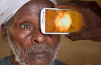
With everyone in the US abuzz about the new iPhone and Apple operating systems, other smartphone advancements are making an even bigger impact worldwide. One of the biggest causes of blindness around the world is due to untreated cataracts and uncorrected refractive errors. Because traditional exam equipment is immobile and very expensive, people in remote rural areas of third world countries rarely receive eye exams or eye care. This is now beginning to change, however, due to advancements with the smartphone and smartphone applications (apps).
A new smartphone app called Peek is revolutionizing the way eye exams are provided in these countries. Using a smartphone and an external clip-on device, Peek can check for cataracts, perform simple vision tests and scan the retina for disease, allowing conditions such as glaucoma, macular degeneration, and diabetic retinopathy to be diagnosed.
Minimal training is required to operate Peek as the information is gathered and then sent to experts around the world for diagnosis. In addition, the GPS data the smartphone and app gather is also very helpful because this information allows for follow-ups and helps other health organizations better target mass treatment campaigns.
Ophthalmologist Thomas Henderson, M.D. offered, “This iPhone application represents a new pathway to bring improved eye care to many people throughout the world. In the United States, I foresee its potential use in emergency room consultations with a physician who is not on site.”
Peek is currently being tested in Kenya, and their team will publish results at the end of their trials in early 2014.
Source: http://singularityhub.com/2013/09/06/will-peeks-mobile-eye-exam-system-take-a-bite-out-of-developing-world-blindness/
Showing posts with label glaucoma. Show all posts
Showing posts with label glaucoma. Show all posts
Wednesday, October 9, 2013
Thursday, August 29, 2013
New Study Ties Early Menopause to Glaucoma
 |
| Women who experience early meno- pause may be at higher risk for dev- eloping glaucoma later in life. |
Glaucoma is caused by fluid accumulating in the eye, which then puts pressure on the eye’s optic nerve. It is a leading cause of blindness in the US, and has no symptoms or pain when it first develops. Medicine, laser treatment, and conventional surgery are used to treat the disease. Ophthalmologist Clayton Falknor, M.D. explained, “While these treatments can help prevent future loss of vision, they do not improve sight already lost from the disease which is why it’s so important to schedule eye exams as you age.”
This study’s result signals that female hormones may protect against the disease. Hormone replacement therapy is thought to reduce fluid pressure in the eye, and researchers of the study also note that hormone levels rise during pregnancy causing fluid pressure in the eye to decrease.
“This study shows the potential value of analysis of large amounts of data. Although I have seen patients for over 30 years, this is not a connection that I have considered. At this time, I am not prepared to prescribe hormone therapy for menopausal women with glaucoma,” said Ophthalmologist Thomas Henderson M.D.
As glaucoma research continues, it’s still very important to schedule comprehensive eye exams as you age to detect any issues and begin treatment, if needed.
Sources:
http://www.tele-management.ca/2013/08/study-links-early-menopause-to-glaucoma-risk/
http://www.nei.nih.gov/health/glaucoma/glaucoma_facts.asp#a
Monday, March 5, 2012
How Optomap Helps Eye Clinic of Austin Provide Better Eye Care
A Word from Dr. Thomas Henderson
Ophthalmology
is the branch of medicine that deals with the health of the eye and the visual
system. As a physician and a patient,
the more I can see, the better I can understand, the better the solution to a
problem.
Optomap
is a unique, high definition, digital imaging system which combines scanning lasers
with a specially shaped (ellipsoidal) mirror to create a panoramic 200 degree
image of the retina inside the eye. The
effect of the wide field is like sticking your head inside a doorway and
looking at the walls instead of peeking through a keyhole.
Dr.Melanie Prosise often uses the Optomap as a convenience for her patients so
that she can see most of the retina without having to dilate pupils. Dr. Clayton Falknor and I frequently use it
to document the important medical details of a particular abnormality of the
retina. The most common photographs are
of the optic nerve head for glaucoma, the central retina for dry or wet macular degeneration, the whole retina for diabetic retinopathy, a particular pigmented
“freckle”, and other areas of interest.
It is much better to compare detailed photographs after 3 months to
detect change quickly, or after a number of years to prove hoped for stability,
than to rely upon vague descriptions or drawings in the medical record. We will soon receive an upgrade to the
Optomap image management software that will make this even easier.
The
most important use of the Optomap is to improve care by helping me improve my
communication with the patient. For
example, in “wet” macular degeneration, I can describe how a new blood vessel
has formed under the retina near the line of sight, is leaking blood, causing
scar tissue and possible loss of sight.
It is better if I can make a drawing or use a model. It is the by far the best if I can show a
patient a photograph of their eye so they can see the reality of the blood
under their central retina, causing distortion and threatening their
sight. In this way we can understand the
problem better and develop a better plan together—and ultimately have a better
chance for a better result.
The
next time you are in, we might use the Optomap to help provide you with better
eye care.
Subscribe to:
Posts (Atom)
