Showing posts with label macular degeneration. Show all posts

Wednesday, November 6, 2013

Research Finds New Eye Drops Could Treat Macular Degeneration

New research published in the journal PLos One earlier this month has many sufferers of age-related macular degeneration (AMD) hopeful for the possibility of a new treatment. 

Age-related macular degeneration is an eye disease that gradually destroys the macula, the part of the eye that provides sharp, central vision needed for seeing objects clearly. According to the Centers for Disease Control and Prevention, almost 2 million Americans over the age of 40 have poor vision caused by AMD.

The new findings experimented with a chemical PPADS (short for pyridoxalphosphate-6-azophenyl-2’,4’-disulfonic acid) to repair AMD-related damage to the eye. Researchers at Tufts University in Massachusetts induced tissue damage and blood vessel growth characteristic of AMD in anesthetized mice and then applied PPADS daily, which resulted in the chemical healing the eye damage. 

The positive implication from this is the fact that a topical application of a drug - for example, in the form of an eye drop - could ultimately be used on humans to treat AMD. Previous research sought to show that certain dietary supplements, such as lutein, were effective in reducing the risk of progressing from dry macular degeneration to wet macular degeneration. This new study has many researchers excited for the possibilities of self-administered treatments. 

Ophthalmologist Thomas Henderson, M.D. explained, “If confirmed in humans, this chemical, given as an eye drop, could potentially reduce or eliminate the huge cost of and need for monthly injections into the eye to stabilize wet macular degeneration and preserve vision.”

Because this research is the first of its kind to demonstrate a topical application of a drug treating wet macular degeneration, much more research is now due in order to confirm this study’s findings.

Photo Credit: Ross Toro, myhealthnewsdaily.com 

Wednesday, October 9, 2013

Smartphone Advances Provide Mobile Eye Exams in Third World Countries

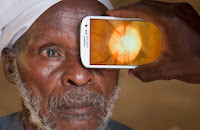
Peek iphone app
With everyone in the US abuzz about the new iPhone and Apple operating systems, other smartphone advancements are making an even bigger impact worldwide. One of the biggest causes of blindness around the world is due to untreated cataracts and uncorrected refractive errors. Because traditional exam equipment is immobile and very expensive, people in remote rural areas of third world countries rarely receive eye exams or eye care. This is now beginning to change, however, due to advancements with the smartphone and smartphone applications (apps).

A new smartphone app called Peek is revolutionizing the way eye exams are provided in these countries. Using a smartphone and an external clip-on device, Peek can check for cataracts, perform simple vision tests and scan the retina for disease, allowing conditions such as glaucoma, macular degeneration, and diabetic retinopathy to be diagnosed.

Minimal training is required to operate Peek as the information is gathered and then sent to experts around the world for diagnosis. In addition, the GPS data the smartphone and app gather is also very helpful because this information allows for follow-ups and helps other health organizations better target mass treatment campaigns.

Ophthalmologist Thomas Henderson, M.D. offered, “This iPhone application represents a new pathway to bring improved eye care to many people throughout the world. In the United States, I foresee its potential use in emergency room consultations with a physician who is not on site.”

Peek is currently being tested in Kenya, and their team will publish results at the end of their trials in early 2014.

Source: http://singularityhub.com/2013/09/06/will-peeks-mobile-eye-exam-system-take-a-bite-out-of-developing-world-blindness/

Wednesday, August 14, 2013

Stem Cell Therapy for Macular Degeneration to Be Tested

macular degenerationResearchers in Japan began recruiting patients on August 1 for a pilot study to determine the possibility of using induced pluripotent stem cell (iPSC) therapy to treat wet-type age-related macular degeneration.

Age-related macular degeneration is a common eye condition for individuals age 50 and older. There are two forms of the disease, dry and wet. The wet form is more severe and is defined by damage to the retinal pigment epithelium (RPE), cells located in the back of the eye.

“Macular degeneration is the leading cause of vision loss in older adults. Currently, there is no way to cure the disease, only slow its progression,” explained ophthalmologist Clayton Falknor, M.D.

In this study, the damaged RPE is removed and researchers then transplant the iSPC-derived RPE cell sheets into the affected area of the eye. Replacing the RPE cells may repair past damage to the patient’s eye, but the issue is finding and using RPE cells that the patient’s immune system does not reject.

Ophthalmologist Thomas Henderson, M.D. said, “If this works as hoped without significant negative side effects, it will revolutionize the therapy of wet macular degeneration.”

If your family has a history of macular degeneration or you believe may have macular degeneration, contact your eye doctor and schedule a comprehensive eye exam. Although research continues in macular degeneration, the current treatment is still slowing the disease and the sooner it can be detected, the bigger impact treatment can have.


SOURCES:
http://www.asianscientist.com/health-medicine/stem-cell-therapy-eye-disease-riken-japan-2013/
http://www.nei.nih.gov/health/maculardegen/armd_facts.asp#1

Monday, December 31, 2012

B49 Omega-3 Fatty Acids and Your Eyesight


Omega-3 fatty acids are definitely a plus in your diet. They are known as a ‘good fat’ for your diet – one that protects the cell membrane and is reported to protect you from certain health issues. One of these health issues, as reported by NPR (National Public Radio), is age-related macular degeneration.

Age-related macular degeneration generally occurs in older adults. This condition results in vision loss in the middle of your line of sight, due to retina damage. According to the article, “Women who ate fish, a prime source of omega-3s, at least once a week were less likely to get age-related macular degeneration compared with women who ate fish just once a month, a new study finds.”

While all of this information is interesting to look at, the article also points out that the study is not complete.
There are always studies being done on vitamins and minerals. It’s important to know what is fact and what is fiction. Doing your homework is essential in preventative medicines, but the most important thing is having a complete eye examination. You eyesight is like a fingerprint – it is unique to you, and what may work for 5 or 10 other people may not necessarily be the best thing for you. If you think you may have an eye disease or your family has history of eye diseases, you should schedule regular eye exams for the best way to keep on top of it.

If you would like to read the article, you can find it here.
Visit our website at www.eyeclinic2020.com to learn more!

Wednesday, June 20, 2012

Press Release - QuantifEye - June 20, 2012

June 20, 2012

Eye Clinic of Austin Implements QuantifEye - Key New Technology for Macular Pigment Measurement

The Importance of Macular Health


A growing number of baby boomers understand the potential effects of aging on their eyesight and are therefore motivated to take action to maintain their independence.  Studies show that today’s “older” Americans are far more active for much longer than previous generations. They stay in the workforce longer and have more hobbies and interests.  Some studies among this group report that loss of independence is actually feared more than death.  So, when older patients are able to drive safely and more confidently, handle bright light easier, read with less strain, and reduce the risk of age related macular degeneration – that is big news.

MACULAR PIGMENT AND MACULAR HEALTH


Macular pigment is composed of photo-protectants and antioxidants.  An apple provides a great example of how antioxidants provide protection. The skin of the apple protects the inside until it is sliced open and exposed to oxygen.  When this occurs, the inside turns brown via a process known as oxidation.  A similar process occurs in our eyes over time as we age.

Dr. Thomas Henderson says, “Macular pigments are like the eye’s ʽinternal sunglassesʼ. Thick, dense, ʽinternal sunglassesʼ block the harmful blue part of the light spectrum that damages one’s eyes across their lifetime. We are pleased to be the first ophthalmology practice in Austin to introduce the QuantifEye non-invasive system for measuring macular pigment optical density.”

INTERVIEW/SEGMENT OP:


Dr. Henderson is available for in-studio talks about this advanced new technology and how it works, as well as all topics related to healthy vision. Please contact Jonna Kieler at 303.447.9192 for further information or scheduling.

Eye Clinic of Austin


Eye Clinic of Austin was founded in January, 1982 by Dr. Thomas Henderson. Dr. Henderson has been involved in all aspects of optical, medical, and surgical patient care, community service through twenty-five years of emergency call at University Medical Center (Brackenridge), drug research and medical lecturing. Dr. Henderson has personally performed over 11,500 cataract and refractive surgery procedures.

Eye Clinic of Austin offers refractive procedures such as blade-free, all-laser LASIK and Intraocular Lens-based procedures such as cataract surgery. Eye Clinic of Austin also provides general eye care such as medical, laser and surgical glaucoma management, diabetic eye care, dry eye relief, as well as a full-service optical shop, and routine and specialty contact lens care and fitting. The doctors at Eye Clinic of Austin include Thomas T. Henderson, MD, FACS, Clayton L. Falknor, MD, and Melanie Prosise, OD.

Monday, March 5, 2012

How Optomap Helps Eye Clinic of Austin Provide Better Eye Care


Ophthalmology is the branch of medicine that deals with the health of the eye and the visual system.  As a physician and a patient, the more I can see, the better I can understand, the better the solution to a problem.
Optomap is a unique, high definition, digital imaging system which combines scanning lasers with a specially shaped (ellipsoidal) mirror to create a panoramic 200 degree image of the retina inside the eye.  The effect of the wide field is like sticking your head inside a doorway and looking at the walls instead of peeking through a keyhole.

Dr.Melanie Prosise often uses the Optomap as a convenience for her patients so that she can see most of the retina without having to dilate pupils.  Dr. Clayton Falknor and I frequently use it to document the important medical details of a particular abnormality of the retina.  The most common photographs are of the optic nerve head for glaucoma, the central retina for dry or wet macular degeneration, the whole retina for diabetic retinopathy, a particular pigmented “freckle”, and other areas of interest.  It is much better to compare detailed photographs after 3 months to detect change quickly, or after a number of years to prove hoped for stability, than to rely upon vague descriptions or drawings in the medical record.  We will soon receive an upgrade to the Optomap image management software that will make this even easier.

The most important use of the Optomap is to improve care by helping me improve my communication with the patient.  For example, in “wet” macular degeneration, I can describe how a new blood vessel has formed under the retina near the line of sight, is leaking blood, causing scar tissue and possible loss of sight.  It is better if I can make a drawing or use a model.  It is the by far the best if I can show a patient a photograph of their eye so they can see the reality of the blood under their central retina, causing distortion and threatening their sight.  In this way we can understand the problem better and develop a better plan together—and ultimately have a better chance for a better result.

The next time you are in, we might use the Optomap to help provide you with better eye care.